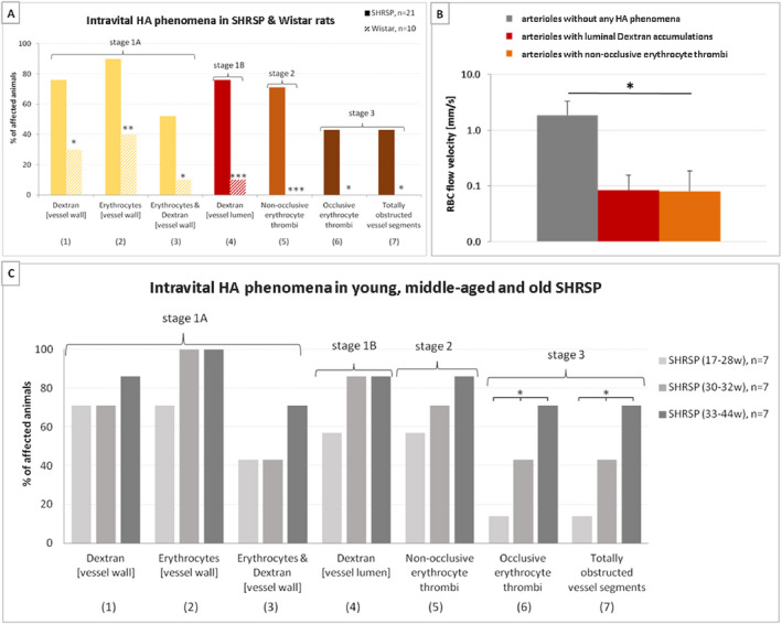
Figure 4.

Frequencies of intravital HA phenomena and RBC flow velocity. A. Frequencies of various small vessel pathology phenomena detected by in vivo imaging (2PM) of the parietal cortex in 21 male SHRSP aged 17–44 weeks (filled bars) and 10 Wistar rats aged 17–35 weeks (striped bars). The yellow bars indicate small vessel wall damage (HA stage 1A), the red ones refer to RBC flow reduction (HA stage 1B), the orange one shows non‐occlusive thrombus formation (HA stage 2), and the brown bars are indicative of occlusive thrombus formation (HA stage 3). Asterisks show significant group differences between SHRSP and Wistar rats (*P < 0.05, ** P < 0.01, *** P < 0.001). B. RBC flow velocity measures revealed lower RBC flow in arterioles with luminal Dextran accumulations (red bar) and significantly reduced RBC flow in arterioles with non‐occlusive erythrocyte thrombi (orange bar) compared to arterioles without any HA phenomena (gray bar). Error bars indicate the standard deviation, *P < 0.05. RBC, red blood cell; RBC flow data refer to the investigation of 8 male SHRSP aged 18–33 weeks; y‐axis—logarithmic scale. C. A separate consideration of young (17–28 weeks, light gray bars), middle‐aged (30–32 weeks, middle gray bars) and old (33–44 weeks, dark gray bars) SHRSP revealed that phenomena (1)–(5) (x‐axis) occurred rather frequently in both age groups while phenomena (6) and (7) showed a significantly higher prevalence in the old compared to the middle‐aged and young animals. * P < 0.05. HA, hypertensive arteriopathy.
