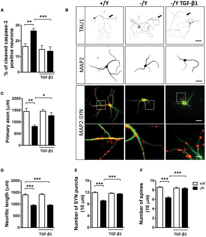
Figure 4.

Effect of treatment with TGF‐β1 on survival and maturation of hippocampal neurons from Cdkl5 KO mice. A. Percentage of cleaved caspase‐3 positive neurons in 4‐day differentiated (DIV4) hippocampal neurons from wild‐type (+/Y n = 5) and Cdkl5 −/Y (n = 5) mice. Hippocampal cultures were treated with TGF‐β1 (1 ng/ml) on day 2 postplating (DIV2). B. Representative images of 10‐day (DIV10) differentiated +/Y and −/Y hippocampal neurons and −/Y hippocampal neurons treated with TGF‐β1 (1 ng/ml), administered on alternate days starting from DIV2, immunopositive for the axon marker TAU1 (upper panel; scale bar = 50 µm, arrows indicate the primary axon), microtubule‐associated protein 2 (MAP2; scale bar = 30 µm), or MAP2 (green) plus synaptophysin (SYN, red). The dotted boxes indicate the regions shown at a higher magnification. Scale bar = 30 μm lower magnification, 2.5 μm higher magnification. C–F. Quantification of the length of the primary axon (C, TAU1‐positive; +/Y = 4, −/Y = 4), the total length of MAP2‐positive neurites (D, +/Y = 6, −/Y = 6), the number of SYN‐immunoreactive puncta per 10 μm in proximal dendrites (E, +/Y = 6, −/Y = 6), and the number of MAP2‐positive spines (F, +/Y = 6, −/Y = 6) from differentiated hippocampal cultures from Cdkl5 +/Y and Cdkl5 −/Y mice treated as in (B). Values are represented as means ± SE. *P < 0.05; **P < 0.01; ***P < 0.001 (Fisher’s LSD after ANOVA).
