
Figure 2.
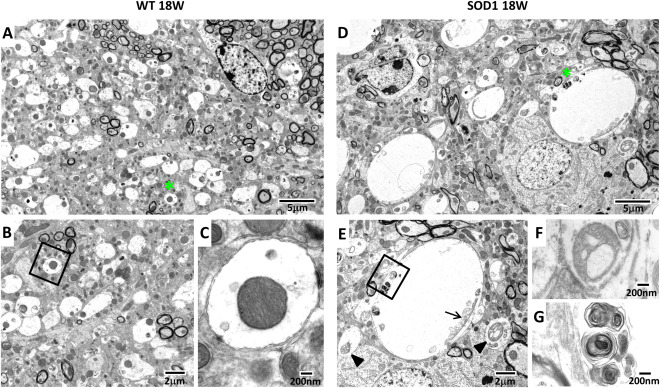
Ultrastructural changes in the AD of SOD1 mice. Representative electron micrographs of the AD from 18 weeks old WT (A–C) and transgenic SOD1 mice (D–G). Tissue from WT exhibits a well preserved tissue structure, including normal sized dendrites (A, asterisks) and mitochondria (B, C). Dendrites from SOD1 mice were expanded (D, asterisk) and mitochondria show various abnormalities, including vacuolation (E, arrowheads) and mitochondrial remnants (E, arrow). Higher magnifications of E show a vacuolated mitochondrion (F) and MLBs (G).