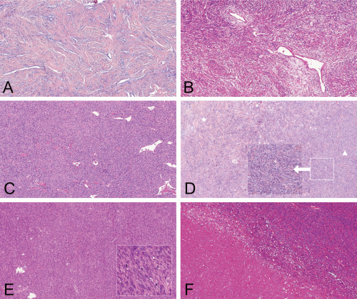
Figure 1.

Histological features and grading of meningeal SFT/HPC. A. MGS grade I: low cellularity and plenty of intervening collagen (“classic fibrous phenotype”), mitotic activity < 5/HPF. (H&E, 100×). B. MGS grade I: variable cellularity (intervening collagen is still present between most of the cells: “classic fibrous phenotype”), mitotic activity < 5/HPF. (H&E, 100×). C. MGS grade I: hypercellularity (“HPC phenotype”), rare intervening collagen and mitotic activity < 5/HPF (H&E, 100×). D. MGS grade I: The cellularity level of this tumor is hard to define precisely: the left area still has collagen between tumor cells and corresponds to a classic “SFT phenotype” (star), whereas more densely packed cells are present in the right side of the microphotograph: possible “HPC phenotype” (arrowhead)—using the updated version of MGS, which is independent of hypercellularity, this tumor is classified MGS I as it displays a mitotic activity < 5/HPF (H&E, 100×. Inset: focus of possible hypercellularity 200×). E. MGS grade II: mitotic activity ≥ 5/HPF without necrosis (H&E, 100×). Inset: high mitotic activity; 400×). F. MGS grade III: necrosis and mitotic activity ≥ 5/HPF (H&E, 100×).
