
Figure 1.
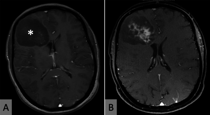
Neuroimaging. Representative axial MRI scans (patient 3) showing a hypointense right frontal expansive mass (*) on T1‐weighted sequences (A). The lesion shows inhomogeneous contrast enhancement (B).
Official websites use .gov
A
.gov website belongs to an official
government organization in the United States.
Secure .gov websites use HTTPS
A lock (
) or https:// means you've safely
connected to the .gov website. Share sensitive
information only on official, secure websites.
Neuroimaging. Representative axial MRI scans (patient 3) showing a hypointense right frontal expansive mass (*) on T1‐weighted sequences (A). The lesion shows inhomogeneous contrast enhancement (B).