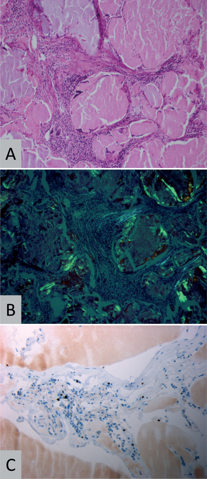
Figure 2.

Histopathology. Representative histopathology (patient 2) showing amyloid deposits surrounded by lymphocytes on H&E staining (A, original magnification 200×) and birefringence under polarized light (B) when stained with Congo red (original magnification 200×). On immunohistochemistry for MiB1/Ki67 (C), few nuclei of lymphoid cells stain positive (original magnification 400×).
