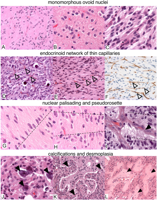
Figure 2.

Histological features of gliomas with FGFR3‐TACC3 fusion. (A–E,G–L) H&E. F. CD34 immunostaining. (A,D,E,F,K,L) 100X. (B,C,G–J) 400X. Recurrent morphological features are: monomorphous ovoid nuclei (A–C), endocrinoid network of thin capillaries (open arrowhead in D–F), nuclear palisading (dash lines in G,H). Tumor cells form pseudorosettes with aligned nuclei (dash line in I) and presence of thin cytoplasmic cell processes between tumor nuclei and vessels (arrowhead in I). J. Microcalcifications (arrowheads). (K,L) Desmoplasia (arrowheads).
