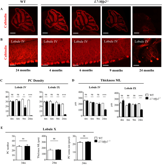
Figure 3.

Anterior to posterior PC degeneration in the absence of MFP2. A. Overview pictures of calbindin stained sagittal sections and B. magnified pictures of anterior lobule IV show progressive PC loss in cerebella of L7‐Mfp2−/− mice. C,D. Quantifications of PC density and ML thickness in lobules IV and IX demonstrate the selective vulnerability of PCs in the anterior cerebellum. E. PCs localized in lobule X are spared from degeneration as their number, soma size and ML thickness are unaltered in both genotypes. N = 4–5 mice per group. Results are displayed as mean ± SEM. NS: not significant, *P < 0.05; ***P < 0.001. Scale bars A: 750 µm, B: 50 µm.
