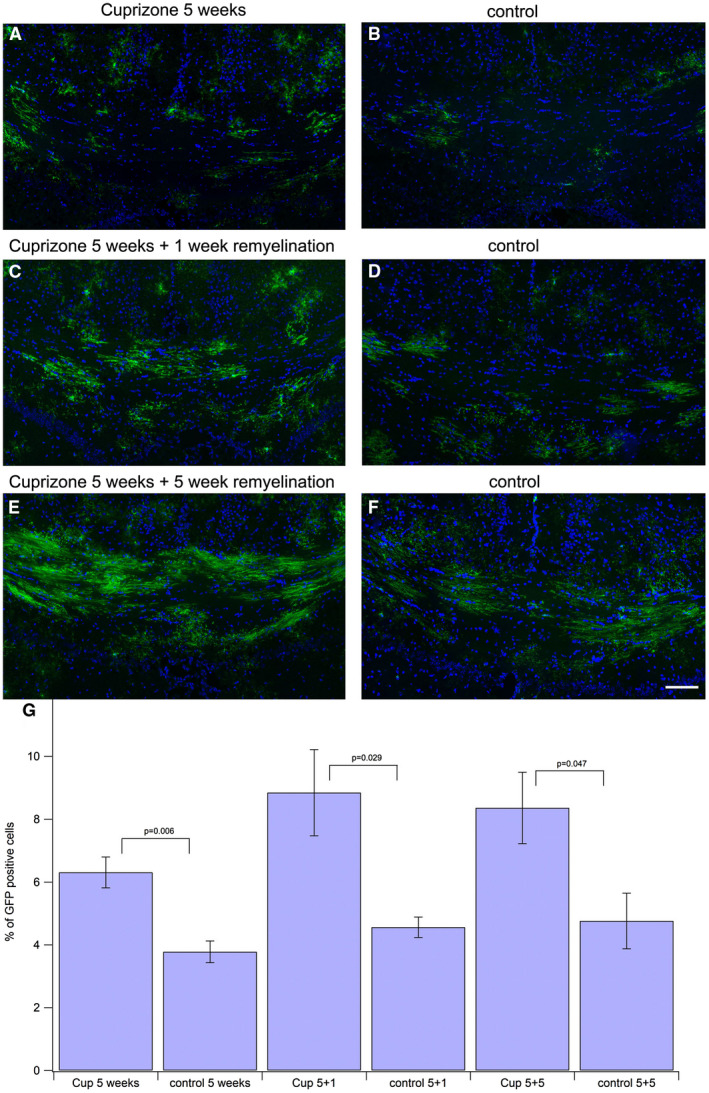
Figure 2.

Amount of GFP‐positive oligodendroglial cells in corpus callosum is enhanced after cuprizone treatment. The GFP‐signal was enhanced by incubating with anti‐GFP antibody and Alexa488 labeled secondary antibody (green). Nuclei were stained with DAPI (blue). All DAPI‐positive nuclei were counted to assess the total number of cells in the corpus callosum. All GFP‐positive oligodendroglial cells were counted to assess the number of newly generated oligodendroglial cells. The ratio of GFP‐positive cells among all cells was calculated and compared to the corresponding controls (tissue taken at same period after tamoxifeninjection). The difference between cuprizone‐treated animals and controls was significant for all groups. Scale bar represents 100 μm. A. 5 weeks of cuprizone feeding, 2 weeks after tamoxifen injection. B. control, 2 weeks after tamoxifen injection. C. 5 weeks of cuprizone feeding plus 1 week of remyelination. Three weeks after tamoxifen injection. D. control, 3 weeks after tamoxifen injection. E. 5 weeks of cuprizone feeding plus 5 weeks of remyelination. Seven weeks after tamoxifen injection F: control, 7 weeks after tamoxifen injection. G. statistical comparison between the treatment groups and the corresponding controls.
