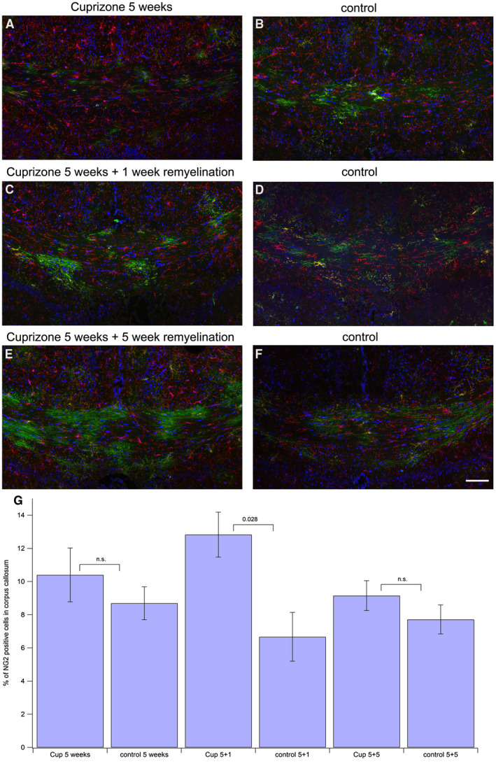
Figure 4.

Assessing NG2‐positive portion of GFP‐expressing oligodendroglial cells. Tissue processing was the same as for Figure 2, except staining for NG2‐positive OPCs was performed with an anti‐NG2 antibody followed by an Alexa633‐labeled secondary antibody (shown in red), GFP (green), DAPI (blue). Scale bar represents 100 μm. A. 5 weeks of cuprizone feeding, 2 weeks after tamoxifen injection. B. control, 2 weeks after tamoxifen injection. C. 5 weeks of cuprizone feeding plus 1 week of remyelination. Three weeks after tamoxifen injection. D. control, 3 weeks after tamoxifen injection. E. 5 weeks of cuprizone feeding plus 5 weeks of remyelination. Seven weeks after tamoxifen injection. F. control, 7 weeks after tamoxifen injection. G. statistical comparison between the treatment groups and the corresponding controls.
