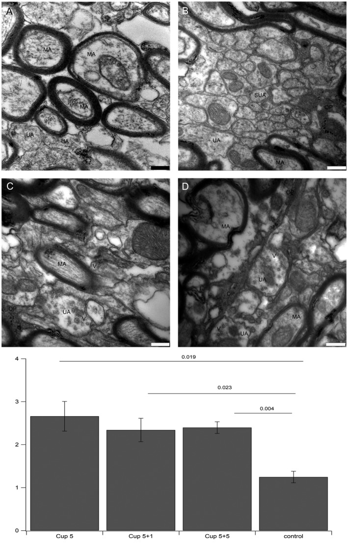
Figure 8.

Ultrastructure of axons. A. myelinated axons in control conditions do not show vesicles. B. small diameter axons after cuprizone treatment do also not show vesicles. C and D. examples for enhanced expression of vesicles in demyelinated axons after cuprizone treatment. E. Statistics: The number of unmyelinated axons containing vesicles per image taken at the same magnification was counted. The differences in all three treatment groups were significantly different from controls. All images were taken at 20.000× fold magnification, bar represents 250 nm. V: vesicle, OP: oligodendroglial protrusion, MA: myelinated axon, UA: unmyelinated axon, SUA: small unmyelinated axon.
