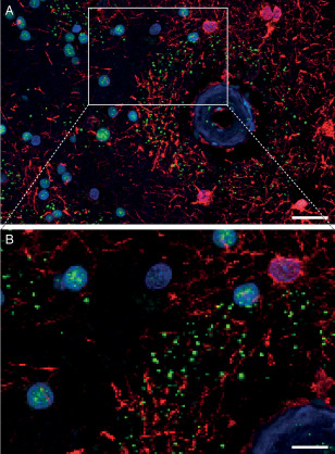
Figure 6.

Perivascular ring of pSMAD2/3 granules limited to the area of GFAP‐positive astrocytes. Immunohistofluorescent double staining with pSMAD2/3 (green), GFAP (red) and nuclei (blue). B detail of A. HCHWA‐D H1 patient‐occipital cortex, merged confocal stack. Scale bar: A. 25 μm; B. 10 μm.
