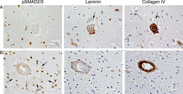
Figure 7.

ECM coarse deposits did not spatially correspond to the pSMAD2/3 granules. Immunohistochemical staining of consecutive slides with pSMAD2/3, laminin and collagen IV antibodies. A. Laminin coarse deposit (and weak collagen IV colocalization; arrows) without presence of pSMAD2/3 granules; HCHWA‐D H5 patient‐occipital cortex. B. pSMAD2/3 perivascular ring of granules (arrows) without perivascular ECM deposits; HCHWA‐D H1 patient‐occipital cortex. Scale bar 25 μm.
