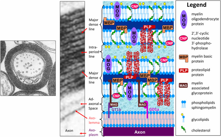
Figure 1.

Electron micrograph of myelinated central nervous system (CNS) tissue at low and high magnifications ([low magnification (left), adapted from Figure 4, 5, 6, 7, originally by Dr. W.T. Norton and Dr. C. S. Raine in Morell P, Quarles RH, Myelin formation, structure, and biochemistry. In Siegel GJ, Agranoff BW, Albers RW, Fisher SK, Uhler MD (editors) Basic Neurochemistry. 6th Edition; 1999. Philadelphia: Lippincott‐Raven; ISBN 0‐397‐51820‐X with permission; high magnification (middle) (adapted from Peters A, Palay SL, Webster H deF. Fine Structure of the Nervous System: The Cells and Their Processes. 1st edition; 1970; page 89, Figure 33, New York: Paul B. Hoeber Inc, with permission from Dr. Alan Peters] depicting the major dense line, which represents the fusion of the cytoplasmic aspects of the oligodendrocyte cell membrane, and the intraperiod line, a potential extracellular space formed by the apposition of the extracellular faces of adjacent oligodendrocyte cell membranes. As shown in the accompanying schematic, the intraperiod line forms a restricted water reservoir, and, thus, is thought to give rise to the short‐T2 component, the signal of which can be displayed anatomically as the myelin water map (see Figure 4, 5, and 6). The oligodendrocyte cell membrane is a bilayer of lipids in which are embedded the major myelin proteins, which include myelin basic protein (MBP), proteolipid protein (PLP), 2′,3′‐cyclic nucleotide 3′‐phosphodiesterase (CNP), myelin oligodendrocyte protein (MOG), and myelin‐associated glycoprotein (MAG). Note, however, that on the inner aspect of the myelin sheath MAG is restricted to the membrane adjacent to the adaxonal space, which it spans to bind the myelin sheath to its axolemmal ganglioside receptors, GD1a and GT1b. The exact position of some of the components of myelin shown in this schematic has not been determined.
