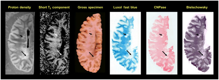
Figure 3.

58‐year‐old male with a 34‐year history of secondary progressive multiple sclerosis (MS) with clinical evidence of optic, cerebellar and spinal involvement. Note the large, irregular lesion in the periventricular occipital white matter, which appears as an area of increased signal on the proton density scan, an area of absent signal on the myelin water/short‐T2 component distribution, and an area of gray discoloration of the white matter in the gross photograph. A band of reduced signal is seen in the lesion on both scans and correlates with the gross appearance (long arrows). More rostrally, several smaller lesions are evident (short arrows), which appear as areas of reduced intensity on the short‐T2 component image. The Luxol fast blue and 2′,3′‐cyclic nucleotide 3′‐phosphohydrolase (CNPase) stains show absence of myelin in most regions of the large periventricular occipital lesion. The Bielschowsky stain for axons is reduced in the lesions but not to the degree of the myelin stains. The faint band detected by the short‐T2 distribution component and the proton density scan is particularly evident on the CNPase stain (long arrows). (Moore, G.R.W., Leung, E., MacKay, A.L., Vavasour, I.M., Whittall, K.P., Cover, K.S., Li, D.K., Hashimoto, S.A., Oger, J., Sprinkle, T.J., Paty, D.W. A pathology‐MRI study of the short‐T2 component in formalin‐fixed multiple sclerosis brain. Neurology 2000;55( 10 ):1506–1510. Figure 1 . Published by The American Academy of Neurology, with permission. http://n.neurology.org/content/55/10/1506.long)
