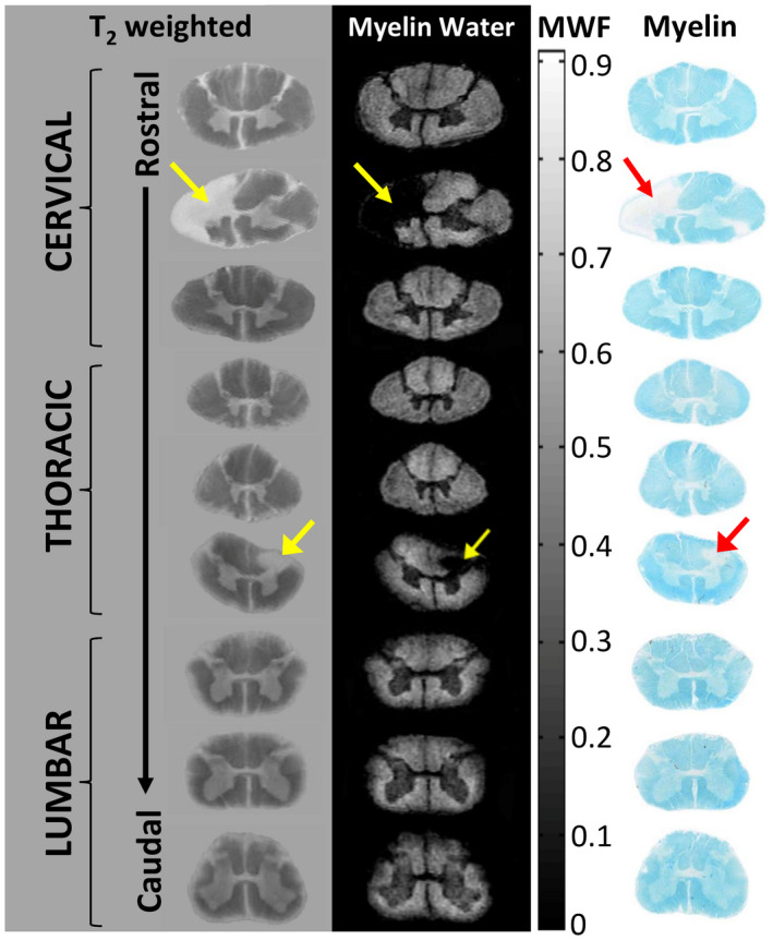
Figure 6.

Magnetic resonance imaging (MRI) and corresponding histology from a formalin‐fixed multiple sclerosis (MS) spinal cord. Cervical, thoracic, and lumbar regions show anatomical variation in myelin with white matter showing increased myelin water relative to the central gray matter butterfly. MS lesions (arrows) demonstrate myelin water loss. Staining for myelin (Luxol Fast Blue) demonstrates excellent correspondence between MRI and histology. (adapted from Figure 1 a, Laule, C., Yung, A., Pavolva, V., Bohnet, B., Kozlowski, P., Hashimoto, S.A., Yip, S., Li, D.K., Moore, G.R.W. High‐resolution myelin water imaging in post‐mortem multiple sclerosis spinal cord: A case report. Multiple Sclerosis Oct 22 2016, 1485–1489, published by SAGE Publications).
