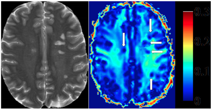
Figure 7.

Heat map of myelin water fraction. Left side: T2 weighted image of a Multiple Sclerosis (MS) patient. Right side: heat map of a myelin water image (MWI). T2‐hyperintense MS‐lesions show clear reductions of myelin water fraction (MWF) (white arrows, right side). (Faizy TD, Thaler C, Kumar D, Sedlacik J, Broocks G, Grosser M, Stellmann J‐P, Heesen C, Fiehler J, Siemonsen S (2016) Heterogeneity of Multiple Sclerosis Lesions in Multislice Myelin Water Imaging. PLoS ONE 11( 3 ): e0151496. https://doi.org/10.1371/journal.pone.0151496 , Figure 2).
