
Figure 2.
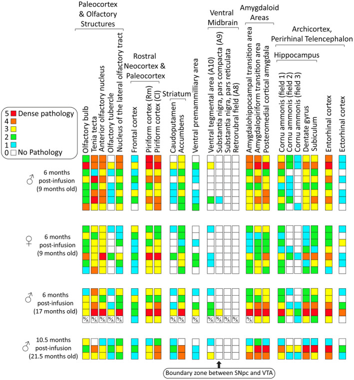
Heat map of α‐synucleinopathy following preformed α‐synuclein fibril injections in the mouse OB/AON. Qualitative assessments of the density of pSer129+ inclusions in the indicated brain regions of all fibril‐infused animals that survived until the planned assay time (see Methods). Each row represents one animal. A blinded investigator systematically examined the entire 1‐in‐5 series of sagittal brain sections of the ipsilateral, right hemisphere following immunostaining of pathologically phosphorylated α‐synuclein with the rabbit monoclonal EP1536Y pSer129 antibody (see Table S1). Boxes with a N/A label denote regions that were unavailable (see Methods). Brain regions with pSer129+ immunolabeling are arranged rostrocaudally and/or by anatomical/functional groupings. Most of the midbrain pathology was confined to the ventral tegmental area; only two male animals exhibited pSer129+ inclusions at the boundary zone between the substantia nigra and the ventral tegmental area at 6 months post‐infusion. Abbreviations are defined in Table S2. https://dsc.duq.edu/pharmacology/ or https://www.dropbox.com/sh/a6r5ylg1tco6trm/AAC9Mb2gWuP29ABbmdGuNoaJa?dl=0