
Figure 4.
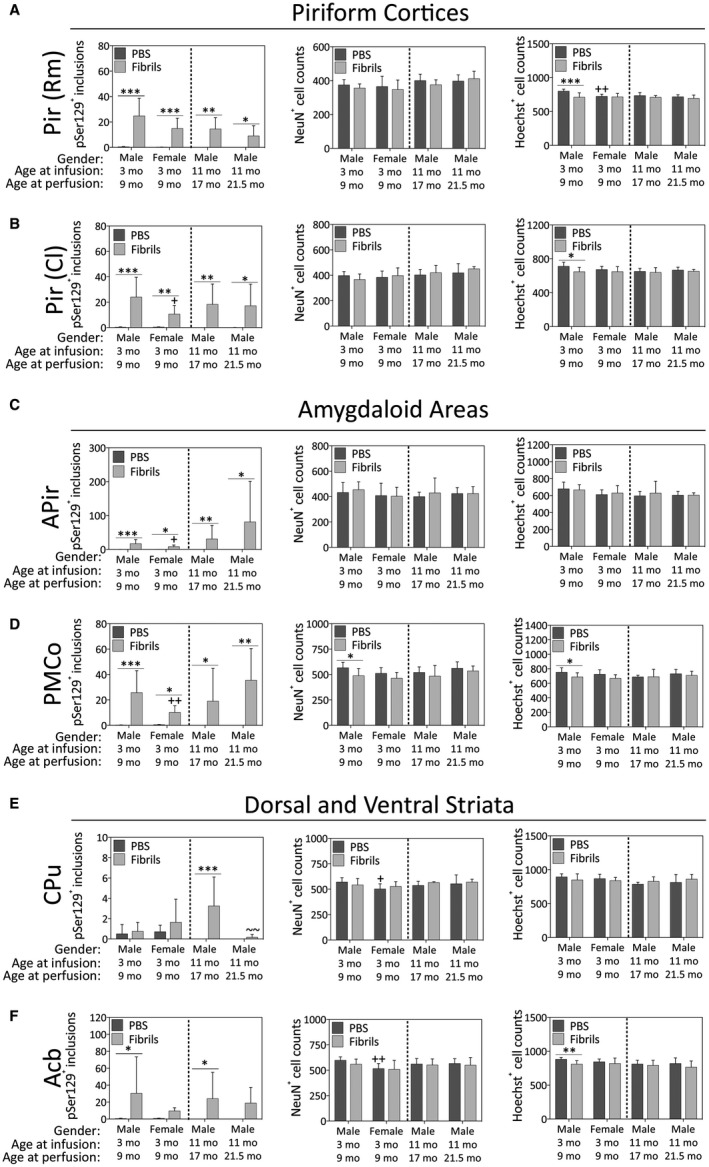
Impact of α‐synuclein fibril infusions in the mouse OB/AON on pSer129+ inclusion counts, NeuN+ neuron counts and Hoechst+ cell numbers in the paleocortices, amygdaloid complex, and the dorsal and ventral striata. Mice were infused in the right olfactory bulb/anterior olfactory nucleus with either preformed α‐synuclein fibrils (5 μg/1 μL) or an equivalent volume of PBS (1 μL). A blinded observer manually counted the number of pSer129+ structures (monoclonal 81A pSer129 Ab; see Table S1) per field of view (200× magnification) and used cellSens software to count the number of NeuN+ and Hoechst+ nuclei in the rostromedial piriform cortex (Pir Rm; A), caudolateral piriform cortex (Pir Cl; B), amygdalopiriform transition area (APir; C), posteromedial cortical amygdala (PMCo; D), caudoputamen (CPu; E) and nucleus accumbens (Acb; F). Shown are the mean and SD of raw, unnormalized data. N = 3–8 mice per group (see Methods and Figure 1 for animal numbers). Two‐way ANOVAs were followed by the Bonferroni post hoc correction. *P ≤ 0.05, **P ≤ 0.01, ***P ≤ 0.001 PBS vs. fibrils; +P ≤ 0.05, ++P ≤ 0.01 vs. 3–9 month males; ~~P ≤ 0.01 vs. 11–17 month males. Abbreviations are defined in Table S2.