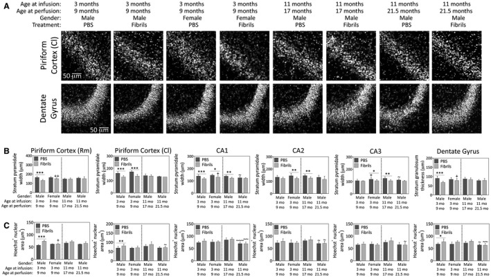
Figure 8.

Preformed α‐synuclein fibril injections in the OB/AON induce atrophy of pyramidal and granule cell layers of the limbic allocortex in young male mice. Mice were infused in the right olfactory bulb/anterior olfactory nucleus with either preformed α‐synuclein fibrils (5 μg/1 μL) or an equivalent volume of PBS (1 μL). A blinded observer employed the “arbitrary line” tool in cellSens software to measure the width of the compact Hoechst+ pyramidal cell layer in the rostromedial piriform cortex, caudolateral piriform cortex, CA1, CA2 and CA3 of the hippocampus, or the stratum granulosum of the dentate gyrus. Representative grayscale micrographs for the caudolateral piriform cortex and dentate gyrus are displayed in panel (A) and the quantifications of pyramidal and granule cell layers width (B) and Hoechst+ nuclear area (C) are graphed below. Shown are the mean and SD of raw, unnormalized data. N = 3–8 mice per group (see Methods section and Figure 1 for animal numbers). Two‐way ANOVAs were followed by the Bonferroni post hoc correction. *P ≤ 0.05, **P ≤ 0.01, ***P ≤ 0.001 PBS vs. fibrils; +P ≤ 0.05, ++P ≤ 0.01, +++P ≤ 0.001 vs. 3–9 month males; ~P ≤ 0.05, ~~P ≤ 0.01, ~~~P ≤ 0.001 vs. 11–17 month males. Abbreviations are defined in Table S2. To view the original, higher resolution Adobe Illustrator or EPS files, please link to https://dsc.duq.edu/pharmacology/ or https://www.dropbox.com/sh/a6r5ylg1tco6trm/AAC9Mb2gWuP29ABbmdGuNoaJa?dl=0
