
Figure 9.
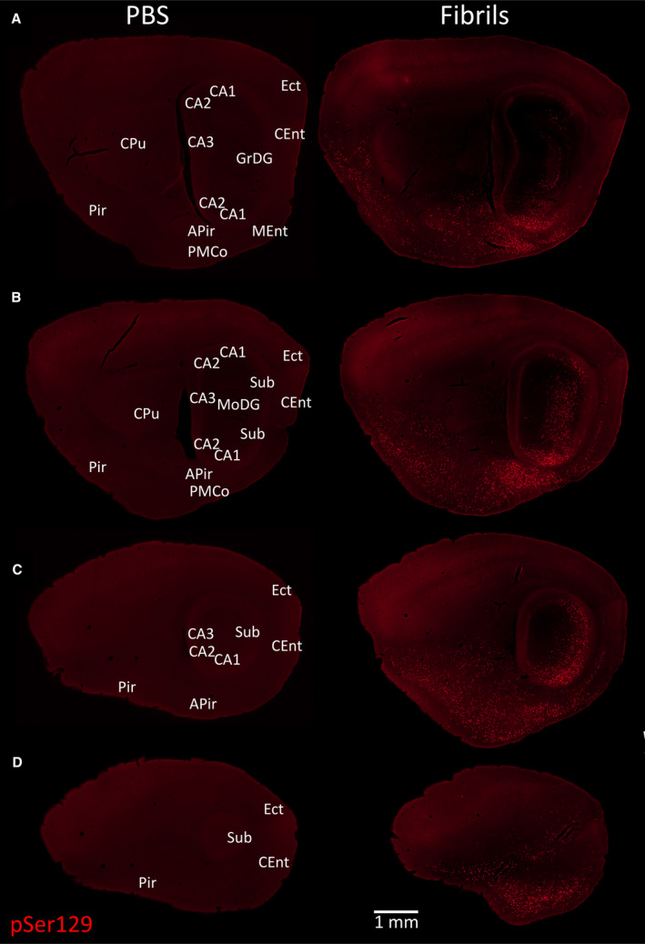
α‐synucleinopathy remains centered in the limbic allocortex 6 months following infusions of preformed α‐synuclein fibrils in the OB/AON. A series of stitched images of sagittal brain sections from two 17‐month‐old animals sacrificed 6 months after infusion of 1 µL PBS (left) or 5 µg/1 µL fibrils (right) into the OB/AON. All sections were stained in parallel with the monoclonal rabbit EP1536Y pSer129 antibody for pathologically phosphorylated α‐synuclein (see Table S1). Anatomical labels are based on cytoarchitectonic details provided by the Hoechst nuclear marker (not shown). All sections were processed in parallel and photographed at the same exposure and intensity scaling. Abbreviations are defined in Table S2. To view the original, higher resolution Adobe Illustrator or EPS files, please link to https://dsc.duq.edu/pharmacology/ or https://www.dropbox.com/sh/a6r5ylg1tco6trm/AAC9Mb2gWuP29ABbmdGuNoaJa?dl=0