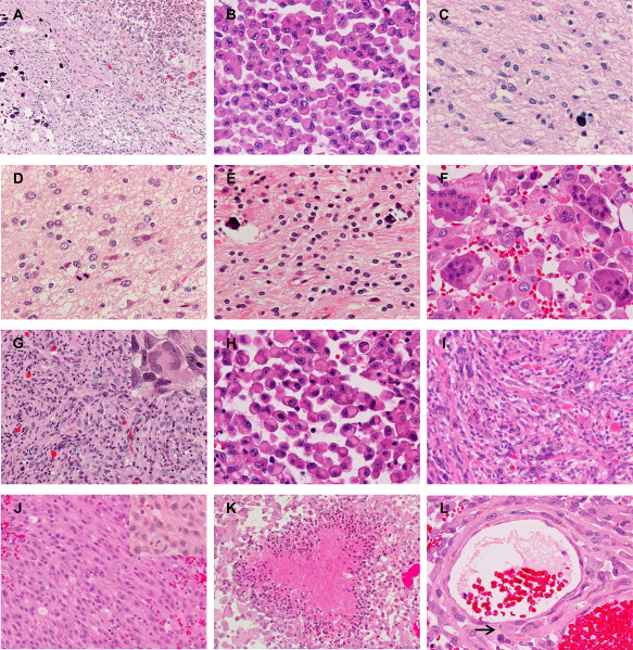
Figure 1.

Microscopic appearance. A–C. Epithelioid glioblastoma (E‐GBM) and diffuse astrocytoma (DA)‐like components in case 9. The interface between the E‐GBM (top right) and DA‐like (bottom left) components is relatively sharp (A). Note the calcification in the DA‐like component (A,C). The E‐GBM area is composed of monotonous, discohesive round cells with laterally positioned nuclei and eosinophilic cytoplasm (B). The DA‐like component exhibits mild cellular proliferation of well‐differentiated neoplastic fibrillary astrocytes (C). D. The anaplastic astrocytoma‐like component in case 4, presenting 4 mitoses per 10 high‐power fields. E,F. The oligoastrocytoma‐like component with calcification (E) and osteoclast‐like giant cells intermingled with epithelioid tumor cells (F) in case 8. G. The PXA‐like component in case 6 shows a fascicular arrangement of spindle‐shaped cells with some multinucleated pleomorphic cells (inset). H,I. E‐GBM (H) and its precursor PXA lesion (I) in case 2. The PXA shows numerous eosinophilic granular bodies (I). J. A component with anaplastic spindle‐shaped cells with monotonous nuclei and thick processes (case 12). Frequent mitosis is seen (inset). K. Palisading necrosis in the E‐GBM component (case 10). L. Epithelioid tumor cells invade the vascular wall in the subarachnoid space (case 6). The arrow indicates a tumor cell right beneath the endothelium. Original magnification: A, x100; G, I–K, x200; B–F, H, L inset in G and J, x400.
