
Figure 2.
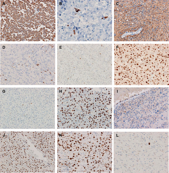
Immunohistochemistry. A. Vimentin is diffusely and strongly positive in E‐GBM (case 9). B,C. A limited number of epithelioid cells with a few cytoplasmic processes are positive for GFAP (B), and the DA‐like area is diffusely positive (C) in case 7. D. Anaplastic spindle‐shaped cells are sparsely immunostained with GFAP (case 5). E,F. The extent and intensity of nuclear Olig2 staining is variable in the E‐GBM components; few and moderate in case 3 (E), whereas diffuse and strong in case 12 (F). G,H. The E‐GBM area has sparse and weak p53 immunoreactivity in case 3 (G), but diffuse and strong in case 13 (H). I. The loss of ATRX nuclear expression in case 4 (E‐GBM area). J. The nuclear staining of INI1 is retained (E‐GBM area of case 3). K,L. The MIB‐1 labeling index is approximately 60% in the E‐GBM component (K) and less than 1% in the DA‐like component (L) in case 3. Original magnification: A, C–L, x200; B, x400.