Figure 3.

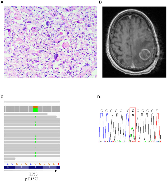
Histology and radiologic feature of a gcGBM from a 42‐year‐old male with TP53 mutation. A. H&E section and B. T1‐enhanced images of the tumor. C. TP53 mutation was visualized with IGV and D. validated by Sanger sequencing.

Histology and radiologic feature of a gcGBM from a 42‐year‐old male with TP53 mutation. A. H&E section and B. T1‐enhanced images of the tumor. C. TP53 mutation was visualized with IGV and D. validated by Sanger sequencing.