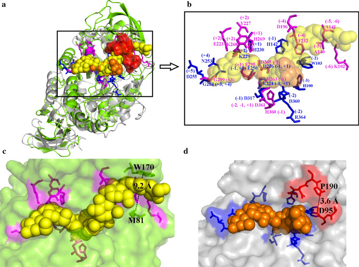
Fig. 6.
Three-dimensional structure alignment of NFAmy13B and NFAmy13A. The overall structure of NFAmy13B (green) and NFAmy13A (gray) were constructed using PDB 1WPC and 3KWX, and showed in cartoon style (a); The substrate-binding sites of NFAmy13B and NFAmy13A are shown as magenta and blue sticks, respectively (b); The surface presentation showed the shape of the pocket, and yellow and orange spheres represent the substrates binding with NFAmy13B (c) and NFAmy13A (d), respectively. Red sticks and surface area indicate the block region of NFAmy13A compared with NFAmy13B

