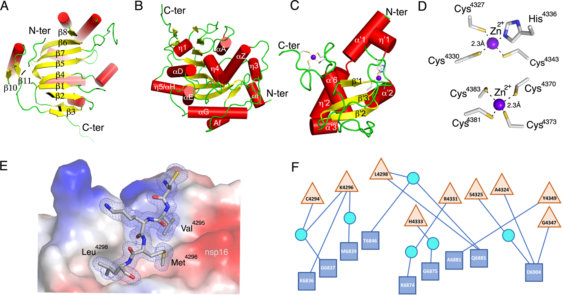
Fig. 2. Detailed representation of nsp16, nsp10, and the heterodimer interface.

(A to C) Cartoon representations of two views of nsp16 featuring the canonical β-sheet (A) and overall secondary structure of nsp16 (B) and nsp10 (C). α-helices are shown as red cylinders, β-strands as yellow arrows, loops as green strands, and zinc ions as purple spheres. (D) Close-up view of the two Zn2+ binding sites in nsp10. (E) Interaction of Cys4294-Leu4298 (sequence CVKML, grey sticks) from nsp10 with the hydrophobic surface of nsp16 (colored by electrostatic potential). Oxygen, red sticks; nitrogen, blue sticks; sulfur, yellow sticks. (F) Schematic representation of residues from nsp16 (blue squares) and nsp10 (tan triangles) that interact through hydrogen bonds, represented as lines. Some interactions are mediated by water molecule (cyan circles). For panels A to D, structural representations are based on the structure of the nsp16-nsp10 complex with m7GpppA and SAM (PDB code 6WVN). E and F are based on the structure of the nsp16-nsp10 complex with SAM in the small unit crystal form (PDB code 6W4H).
