Fig. 3.
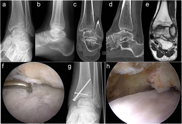
Eighteen-year-old male patient (case 11) with a posttraumatic osteochondral lesion of the medial talus, which was treated with primary combined matrix-associated autologous chondrocyte implantation (MACI) via medial malleolar osteotomy with autologous bone grafting (ABG) from the ipsilateral iliac crest; preoperative antero-posterior (a) and lateral (b) radiographs; preoperative computed tomography scans in the coronary (c) and sagittal (d) plane; preoperative magnetic resonance image in the coronary plane (e); arthroscopic image at the time of chondrocyte harvesting (f) showing the cartilage damage at the medial talus and the status after debridement of a ventral tibial osteophyte; antero-posterior radiograph (g) after MACI/ABG showing screw fixation of the medial malleolar osteotomy; arthroscopic image at the time of hardware removal (h) 23 months after MACI/ABG showing the cartilage surface at the medial talus

