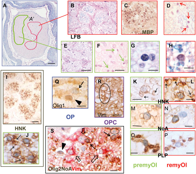
Figure 4.

Morphology and immunohistochemistry of neuroglial cells in totally demyelinated multiple sclerosis optic nerve (MSON) with demyelination lesion type A (DMA). A, B, D–H. Luxol fast blue (LFB)‐stained sections at overview (A) to high power magnification show the presence of cells with small round nuclei (arrows in D, F) that are closely associated with thin (MBP +, C) myelin sheaths in some fascicles (outlined in red on overview, A and boxed in red; B–D, H) but not in others (outlined in green on overview, A and boxed in green; E–G). I–L. Immunohistochemistry with HNK labeled almost all of these cells, resulting in extensive staining throughout the demyelinated fascicles (I). J–L. At high magnification, HNK + staining was seen in two distinct patterns. J, K. In the majority premyOls (individual images boxed in green), radiating processes were seen extending out from perinuclear cell bodies to sometimes end in periaxonal cuffs (arrows), whereas remyOls (individual images boxed in red) lacked visible processes such that their perinuclear positivity was separated from nearby periaxonal rings (arrows in L). Further analysis showed a similar discriminative pattern with the markers NoA (M, N) and PLP (O, P). C. Immunohistochemistry with MBP was restricted to the thin myelin sheaths of remyOls. Q–R. In addition to premyOls and remyOls, antigenic phenotyping identified occasional OPC and OP. R. Following the SIMPLE (Sequential Immunoperoxidase Labeling and Erasing) protocol, an OPC nucleus at the top of the outlined example is faintly stained with haematoxylin remaining from the previous imaging stage and encircled by Vim+ cytoplasm that extends distally to wrap around nearby axons, (Vim preceding HNK and final counterstain). Q. NG2 cells/OP were identifiable by nuclear Olig1 expression (arrowhead) in contrast to premyOls or remyOls that had weak cytoplasmic staining (arrow). S. Dual staining identified multiple neuroglial cell types simultaneously within a single preparation. Olig2 combined with NoA (brown) and contrasted with Vim (red), separated premyOls or remyOls with small round (weakly) Olig2+ nuclei and NoA + cytoplasm (Olig2+/NoA +/Vim−; arrows) from NG2 cells/OP that had intense Olig2+ nuclei but no cytoplasmic staining (Olig2+/NoA −/Vim−; arrowhead), astrocytes with large nuclei and coarse straight processes (Olig2−/NoA −/Vim+; open arrows) and immuno‐negative microglia (Olig2−/NoA −/Vim−; circled). Scale bars = 1 mm (A); 50 μm (E, I); 20 μm (F); 10 μm (P–R); 5 μm (G, H, J–S).
