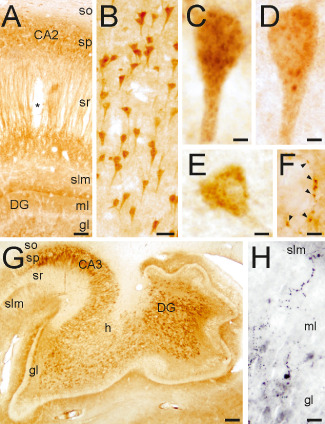
Figure 2.

CPE and SgIII protein expression in the hippocampus. (A) CPE distribution in the hippocampus. (B) Pyramidal cell bodies labeled for SgIII. (C,D) Granule‐like compartments stained for SgIII (C) and CPE (D) in CA1 pyramidal neurons. (E) A CPE‐positive interneuron in the stratum oriens. (F) CPE‐labeled terminal‐like buttons on a negative soma in the hilus. (G) Pattern of SgIII immunolabeling in CA3 and the dentate gyrus. (H) SgIII varicose fibers in the inner portion of the molecular layer of the dentate gyrus. Scale bars in μm: A, 150; B, 50; C‐F, 5; G, 1000, H, 25. Abbreviations: so = stratum oriens; sp = stratum pyramidale; sr = stratum radiatum; slm = stratum lacunosum‐moleculare; ml = molecular layer; gl = granular layer; h = hilus; DG = dentate gyrus.
