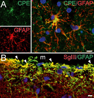
Figure 3.

Astroglial‐identified cells express CPE and SgIII in the cerebral cortex. (A, B) Confocal images illustrating double immunofluorescence of CPE and SgIII with GFAP in the white matter (A) and the upper layers of the parietal cortex (B). Note the location of CPE in the perinuclear area and processes of astrocytes. In B, SgIII decorates punctuate structures and thin processes exhibiting GFAP. Arrowheads indicate yellow color in merge images. Nuclei are in blue color. Scale bar in μm: A, 10; B, 5. M = meninge.
