Figure 4.

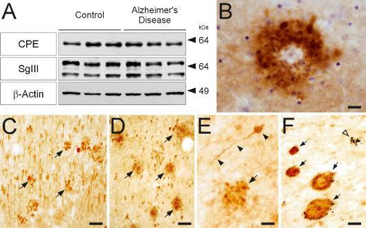
CPE and SgIII are accumulated in senile plaques of AD patients. (A) Western blots showing protein levels of CPE and SgIII in homogenates of hippocampus from AD patients and age‐matched controls. Densitometric analyses revealed no significant differences among groups in protein expression levels, normalized to β‐actin (P > 0.05). The mobility of molecular mass markers (in kDa) is indicated. (B–F) Aberrant accumulation of SgIII (B,D,F) and CPE (C,E) are consistently detected in AD plaques of different areas. (B) SgIII‐labeled corona plaque in upper layers of the neocortex, Nissl counterstaining in blue. (C,D) Arrows indicate numerous plaques in the CA1 region of the hippocampus. (E) CPE‐positive dystrophic neurite (arrowheads) and a plaque (arrow) in the white matter. (F) Double immunostaining against SgIII (brown) and Aβ (blue) in the hippocampus showing four double‐labeled senile plaques (arrows) and one lacking SgIII. Scale bar in μm: B, 20; C,D and F, 50; E, 15.
