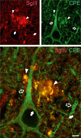
Figure 5.

Colocalization of SgIII with CPE in AD senile plaques. Confocal images showing double immunofluorescence of SgIII and CPE in layer V of the parietal cortex. Aberrant plaque structures display a high grade of overlapping (yellow color in merge image, arrowheads). Note the differential distribution of SgIII (arrows) and CPE (open arrows) in the neuropil and subcelullar locations around the nucleus. Scale bar: 5 μm.
