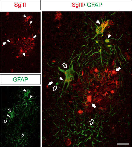
Figure 7.

Increased levels of SgIII in plaque‐surrounding reactive astrocytes. Confocal double immunofluorescence of SgIII and GFAP in the AD hippocampus. Hypertrophied GFAP‐labeled astrocytes that surround an amyloid plaque aberrantly contain high levels of SgIII. Arrows and open arrows indicate single labeling structures. Yellow color in merge image indicates colocalization (arrowheads). Scale bar: 25 μm.
