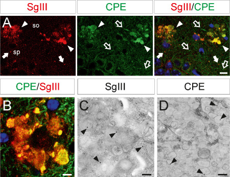
Figure 9.

CPE and SgIII accumulation in dystrophic neurites of transgenic mice. CPE and SgIII colocalization in degenerating neurites in the CA1 region of the hippocampus (upper images) and somatosensorial cortex (lower image) by double confocal immunofluorescence. Blue color labels cell nuclei. Ultrastructural images show immunogold staining of SgIII and CPE in autophagic‐like (left) and enlarged (right) vesicles in cortical dystrophic neurites. Scale bars in μm: A, 10; B, 5; C and D, 0.5. Abbreviations: so = stratum oriens; sp = stratum pyramidale.
