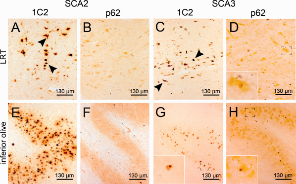
Figure 4.

Aggregates in the lateral reticular nucleus and the inferior olive (A‐D). The lateral reticular nucleus (LRT) of the medulla oblongata displays severe GCS in a SCA2 and a SCA3 case (A,C) (arrowheads), while showing almost no NNI in the SCA2 patient (B) and only a singular NNI in SCA3 (D, insert). (E–H) The inferior olive shows intense GCS in SCA2 (E) and only moderate GCS in SCA3 case (G), while discernable NNI are almost absent in SCA2 but frequently present in SCA3 (G,H). A,C,E,G: 1C2 staining; B,D,F,H: p62 staining; 100 μm PEG embedded sections.
