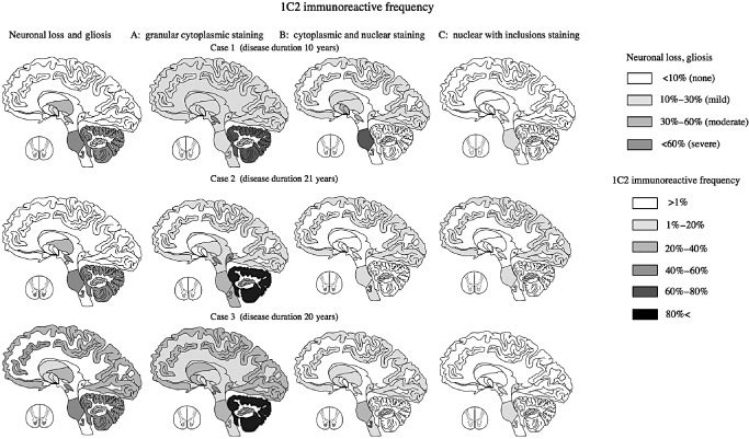
Figure 6.

Distribution according to neuronal loss, gliosis, and 1 C 2‐immunoreactive frequency (1 C2‐IF ) of three labeling patterns in three patients with spinocerebellar ataxia type 2 ( SCA 2). The first pattern (cytoplasmic granular staining) was seen in almost all diffuse lesions in all three SCA2 cases. The second pattern was seen mainly in degenerative lesions (those with neuronal loss and gliosis) except for in the cerebellum. The third pattern was found in severely and mildly degenerative lesions.
