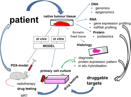
Figure 1.

Craniopharyngioma research facets. Based on the most important source of this research, the patient‐derived native tumor tissue obtained during surgical treatment, molecular analyses reveal druggable targets (gray arrows). Both in vitro and in vivo models are indispensable for the functional analysis of identified targets. Primary cell cultures and patient‐derived xenograft (PDX) models were established from native ACP tissue. The efficacy of drugs could be examined using both models. Because of the limits of cell cultures, gene regulatory events based on the inhibition of a specific target should be verified by histology, for example, utilizing double immunofluorescence to analyze the target, as well as the expression of the identified gene (green arrow). In contrast to cell cultures, the PDX model consistently conserves molecular tumor appearances, as seen in the patient. Therefore, this model seems to be most suitable for translating results into humans. Tumor screening using MRI and the development of radiotherapy protocols, adapted for the clinical course, not only allow the further investigation of mechanisms leading to recurrence even after irradiation, but also drug primed enhancement of radiosensitivity. Using these models, our prospective goal is the identification of patient precision adjuvant therapy (broken arrow).
