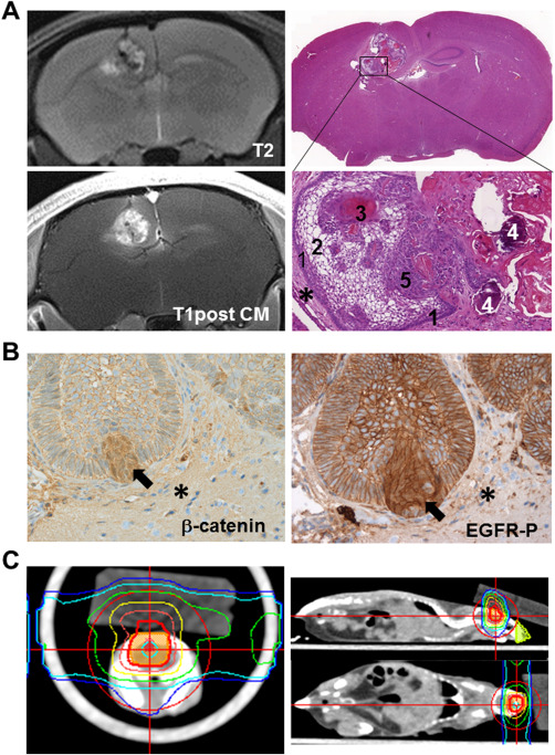
Figure 3.

PDX model. (A) Intracranial lesion of transplanted patient‐derived ACP tissue is detectable on T2‐weigted and after contrast media administration T1‐weigted MR images. Hematoxylin & Eosin staining reveal histological appearance of a tumor transplanted into the mouse brain (asterisk), showing typical histological hallmarks in magnification, for example, palisading cells (1), stellate reticulum cells (2), wet keratin (3), calcification (4) and solid cells (5) of ACP. (B) Xenotransplanted tumors also exhibit digitate tumor protrusions into adjacent brain tissue (asterisk) with beta‐catenin accumulating cells (arrow) also showing EGFR activation (EGFR‐P) with nuclear localization (arrow). (C) In order to evaluate the impact of radiation therapy on ACP tissue, PDX model irradiation planning was performed in accordance with the standard clinical approach. Colored isodose lines correspond to the dose levels shown in the coronal, sagittal and axial view (adapted from Hartmann et al).
