
Figure 4.
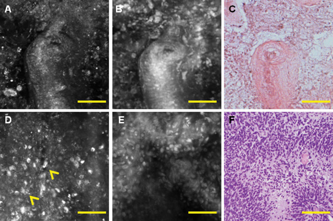
Correlation of histology and multimodal confocal microscopy shows radiation damage with hyalinized blood vessel and necrosis on reflectance (A) and fluorescence images (B) similar to the corresponding haematoxylin and eosin (H&E) (C). Pseudopallisading necrosis, another hallmark feature of glioblastoma (GBM), can be visualized as mirror images of bright necrosis by reflectance (D), dense accumulation of tumor nuclei on fluorescence (E), similar to the corresponding H&E area (F). Scale bar represents 100 μm.