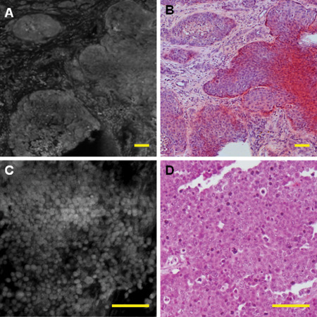
Figure 5.

Metastatic tumors are usually highly cellular on fluorescence and can recapitulate the architecture of their primary tumor, such as adenocarcinoma [A, fluorescence; B, corresponding haematoxylin and eosin (H&E)]. However, some metastatic tumors similar to H&E show only poorly differentiated neoplasm, which needs further workup (C,D). This was an example of metastatic poorly differentiated breast carcinoma. Scale bar represents 100 μm.
