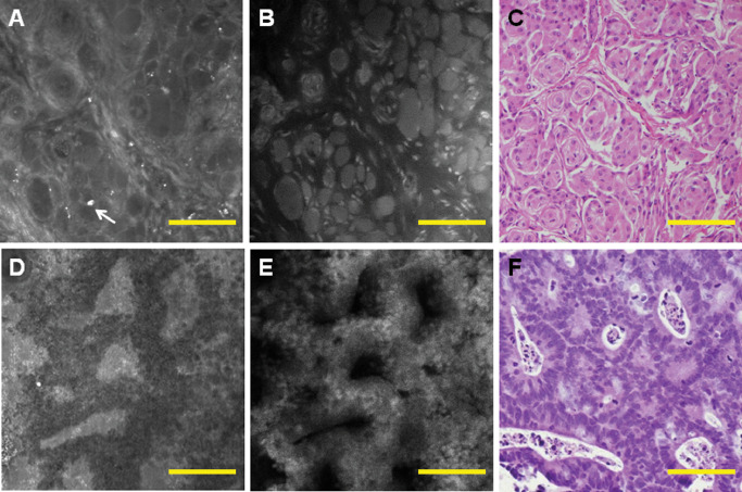
Figure 6.

In nonglial brain tumors, fluorescence and reflectance can complement each other. An example of meningioma shows that the outside region of the whorls is highlighted by reflectance (A), while cell bodies are highlighted by fluorescence (B), and both images closely resemble corresponding haematoxylin and eosin (H&E) (C). Dystrophic calcifications, which are commonly found in meningiomas, are highlighted by reflectance (A, arrow). Adenocarcinoma glands can be visualized well by reflectance (D) and fluorescence (E) and reveal a morphology similar to H&E (F). Scale bar represents 100 μm.
