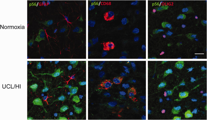
Figure 3.

Immunohistochemical analysis of phospho‐S6 expression in the rat brain at P10 following UCL/HI at P6 compared to normoxic controls. Representative immunohistochemical stains of deep cortical layers of parietal lobe showing increased phospho‐S6 (pS6) expression in neurons of rats 4 days after UCL/HI compared to age and sex matched normoxic controls. There is no pS6 coexpression on astrocytes (GFAP+) or oligodendrocytes (OLIG2+) in neither UCL/HI rats nor normoxic controls. pS6 coexpression is visible on double staining for resting and activated microglia (CD68+) in UCL/HI rats, but not normoxic controls (bar size 50 µm).
