
Figure 1.
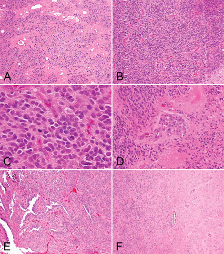
An illustration of selected histological features, including. A. Areas of low cellularity, B. areas of hypercellularity, C. mitoses, D. microvascular proliferation, E. extensive ependymal canals and F. tumor interface with the adjacent brain parenchyma.