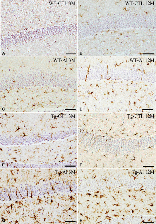
Figure 4.

GFAP‐positive astrocytes in hippocampus in representative tau Tg and WT mice with and without Al treatment. A,B. WT mice without Al treatment (WT‐CTL). At 3 months, only a few, scattered astrocytes were weakly immunopositive for GFAP (A). They were slightly increased in number at 12 months (B). C,D. Al‐treated WT mice (WT‐Al). A moderate number of GFAP‐positive astrocytes were noted at 3 months (C). The number of labeled astrocytes slightly increased with age (D), and it was also larger than that in a WT‐CTL mouse (B). E,F. Tau Tg mice without Al treatment (Tg‐Al). A moderate number of GFAP‐positive astrocytes were seen at 3 months (E), and they increased slightly in number up to 12 months (F). G,H. Al‐treated tau Tg mice (Tg‐Al). Abundant intensely labeled astrocytes were already noted at 3 months (G). The number of labeled astrocytes at 3 months was comparable to that at 12 months (H). GFAP immunohistochemistry. All scale bar = 50 μm.
