Figure 6.

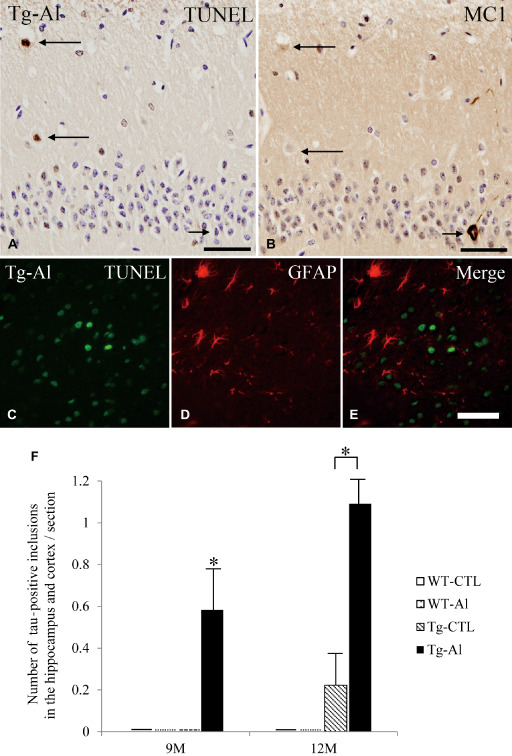
TUNEL‐positive cells in hippocampus, and its relationship to tau‐positive inclusions and GFAP‐positive astrocytes. A. TUNEL‐positive cells (long arrows) in the hippocampus in a 12‐month‐old Tg mouse treated with Al. B. This picture was taken from the identical region of a mirror section of (A) and reversed. TUNEL‐positive cells shown in (A) do not show tau (MC‐1) immunoreactivity (long arrows). Conversely, a neuron containing tau aggregation (short arrow in B) is TUNEL‐negative (short arrow in A). C–E. Double‐labeled immunofluorescence in the hippocampus in 12‐month‐old Tg‐Al mice did not show co‐localization of TUNEL and GFAP immunoreactivities. (A,B) scale bars = 50 μm, (C–E) scale bars = 30 μm. F. Mean numbers of TUNEL‐positive cells in the hippocampus of four groups (ie, WT‐CTL, WT‐Al, Tg‐CTL, Tg‐Al) at 3 months and 12 months. The data at 6 months and 9 months are not shown because no mouse had a TUNEL‐positive cell. The mean number of TUNEL‐positive cells in the hippocampus in Tg‐Al mice was significantly larger than in the other three groups. Statistical analysis were performed by one‐way ANOVA and Tukey post hoc test (***P < 0.001). The error bars represent SEM.
