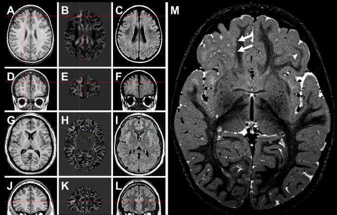
Figure 1.

Neuroimaging findings in MOGHE. A–F. Patient #2 (Table 1). G–L. Patient #13 (Table 1). In the latter patient, the lesion became evident only after morphometric MRI analysis (junction image in 2nd column). 23 Red crosses highlight a suspected lesion in T1 (1st column), and postprocessed junction maps (2nd column). 3rd column: FLAIR sequences at same section planes. M. Three‐year old female patient with band‐like signal alteration at the gray‐white matter junction in right frontal lobe (arrows in T2 weighted turbo echo spin image) and histopathologically confirmed MOGHE.
