Figure 2.

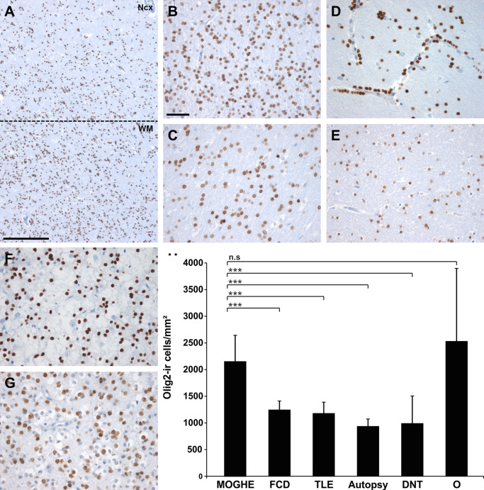
Oligodendroglial cell densities in epilepsy specimens and controls. A. Olig2 immunohistochemistry at the gray (NCx) ‐white matter (WM) junction in MOGHE (dotted line). Scale bar = 500 μm. B–G. higher magnification from white matter regions in different pathology samples and post‐mortem controls (autopsy; E). B. MOGHE; (C) FCD ILAE Type I, (D) temporal lobe epilepsy (TLE) with perivascular clustering of oligodendroglia‐like cells 25, 39; (F) DNT (WHO I°), (G) oligodendroglioma (O; WHO II°). Scale bar in B = 20 μm, applies also to C–G. H. a statistically significant increase was detected for Olig2‐positive cells in MOGHE and oligodendrogliomas (O). *** = P < 0.001, n.s. = not significant.
