
Figure 2.
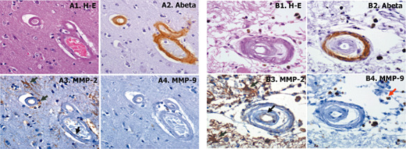
A. Representative example of cerebral amyloid angiopathy (CAA) grade 2; brain vessels that present the media muscle layer replaced by amyloid. B. Representative example of CAA grade 3; vessel with double‐barrel appearance. Serial section were stained with hematoxylin‐eosin (H‐E) staining (A1 and B1), anti‐β‐amyloid (A2 and B2), anti‐matrix metalloproteinase (MMP)‐2 (A3 and B3) and anti‐MMP‐9 (A4 and B4) antibodies. Cellular positive staining is indicated with black arrows for endothelial cells and green arrows for reactive astrocytes 400× magnification.