
Figure 1.
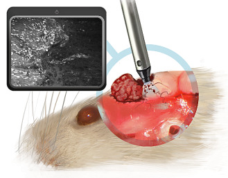
Schematical draft illustrating the relation between mouse brain, tumor and confocal endomicroscopy device in a case of convexity meningioma. The posterior part of the tumor has been removed to scan the tumor bed with the confocal endomicroscopy device and detect invasive meningioma cells along blood vessels in the brain parenchyma.