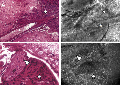
Figure 5.

Identification of intratumoral nerves and vessels through handheld confocal imaging. For each image, H& E‐stained section is displayed on the left panel and confocal image is shown on the right panel. Detection of intratumoral nerves (A) and vessels (B) by handheld confocal imaging. Losange: meningioma; asterisk: blood vessel lumen; arrow head: nerve; star: meningioma.
